A safe, usually comfortable scan that uses sound waves to see the uterus and ovaries. Your scan is read together with your symptoms, so the findings lead straight to a plan.
Sound waves, not radiation.
Over the tummy (abdominal) or through the vagina (transvaginal).
Usually takes 10–20 minutes. It helps find causes of period problems, pelvic pain and fertility concerns.
Abdominal: full bladder. Transvaginal: empty bladder.
Sex determination of the foetus is never done. It is illegal in India.
A pelvic ultrasound (USG pelvis) is a safe, usually comfortable scan that uses sound waves to create images of the uterus, cervix, ovaries, fallopian tubes and bladder. It uses no radiation, so it is safe for women of all ages.
Pelvic ultrasound is often the first test a gynaecologist uses to find the cause of period problems, pain or fertility concerns. At Mannat Clinic, your scan is read together with your symptoms, so the findings lead straight to a plan.
No radiation. Safe at all ages.
Ultrasound uses sound waves only.
The probe is moved over your lower abdomen with a gel. Usually needs a full bladder.
A thin, covered probe is gently placed in the vagina for clearer, closer images. Done with an empty bladder, only with your consent; usually suitable for women who are sexually active.
Checks blood flow to the ovaries or uterus, for example in suspected ovarian torsion.
A series of scans to track egg growth and ovulation when planning pregnancy.
Sometimes both are done in the same visit. This gives a wide view and a close view together.
For many problems, the scan can be done on any day. For some, timing gives clearer answers. Your gynaecologist will tell you the right day when you book.
Drink 4–5 glasses of water about an hour before, and don’t pass urine until the scan.
Empty your bladder just before the scan. No water is needed.
What to expect: the scan usually takes 10–20 minutes. A female attendant is present. Your gynaecologist explains the findings during the same visit. You can eat, drive and go back to work straight after; many women from ITPL, Brookefield and Mahadevapura book an early morning or evening slot.
Your doctor will explain what each finding means for you. Please don’t rely on online searches alone to read your report.
The thickness of the womb lining. Normal values change with your cycle and with menopause.
A uterus larger than usual. It may be due to fibroids, adenomyosis or past pregnancies.
A thin-walled, fluid-filled sac. Many are functional cysts that go away on their own.
Ovaries with many small follicles. This is one feature of PCOS, not a diagnosis on its own.
A small amount is often normal around ovulation. A larger amount may need checking.
If needed, your doctor may advise an MRI, a tube test (HSG or sonosalpingography), hysteroscopy or laparoscopy.
Go to a hospital emergency department straight away if you have sudden, severe pain on one side of the lower tummy · a positive pregnancy test with pain, bleeding or fainting · heavy bleeding soaking a pad every hour · pelvic pain with high fever or vomiting. These may be signs of ovarian torsion, ectopic pregnancy or infection, which need emergency care.
Sex determination of the foetus is not done at this clinic. It is illegal and a punishable offence under the Pre-Conception and Pre-Natal Diagnostic Techniques (PCPNDT) Act, 1994.
PCPNDT Registration No.: [add registration number]A short drive from Kundalahalli, Hoodi, Marathahalli and Varthur. Ultrasound is done only on the advice of a registered medical practitioner.
Ultrasound interpreted alongside your symptoms by a gynaecologist
Diagnosis and treatment planning in the same visit
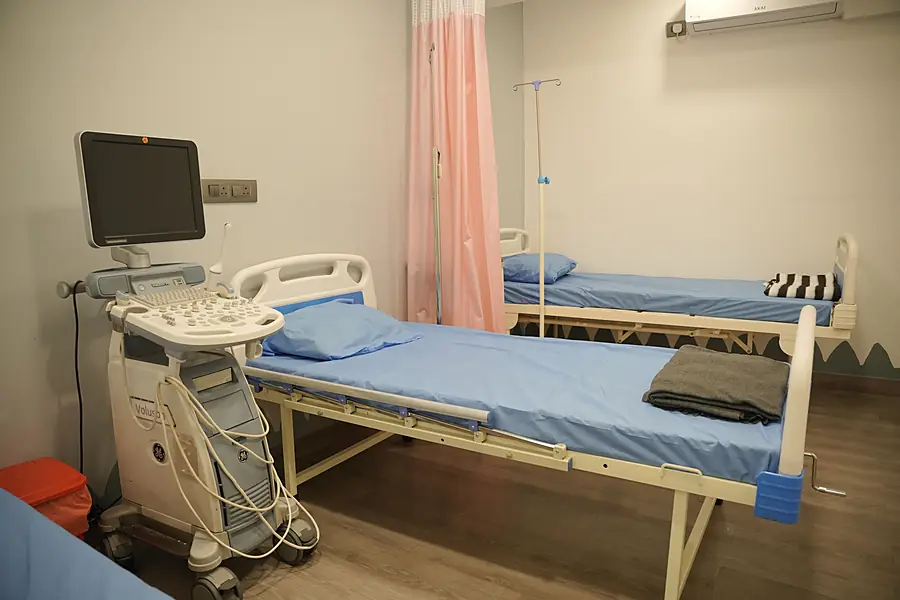
Comfortable, private scanning room
Female attendant present during the scan
Open 7 days, 8 AM – 8 PM, near Kundalahalli Signal
About pelvic ultrasound scans.
Most women feel only mild pressure. Tell us if you feel uncomfortable and we will stop.
Yes, a scan can be done during periods. For some conditions, your doctor may advise a specific day of your cycle.
Yes. It uses sound waves, not radiation.
Ultrasound can show polycystic ovaries, but PCOS is diagnosed together with your symptoms and blood tests.
Usually on the same day.
A TVS is done only with your consent. If you are not sexually active, an abdominal scan is usually done instead.
For an abdominal scan, drink 4–5 glasses about an hour before. No water is needed for a transvaginal scan.
Ultrasound can show changes that need a closer look. A diagnosis needs further tests, such as a biopsy or blood tests.
No matching question. Please call or WhatsApp us and we'll help.
Get clear answers about your pelvic health. Ultrasound is done only on the advice of a registered medical practitioner.